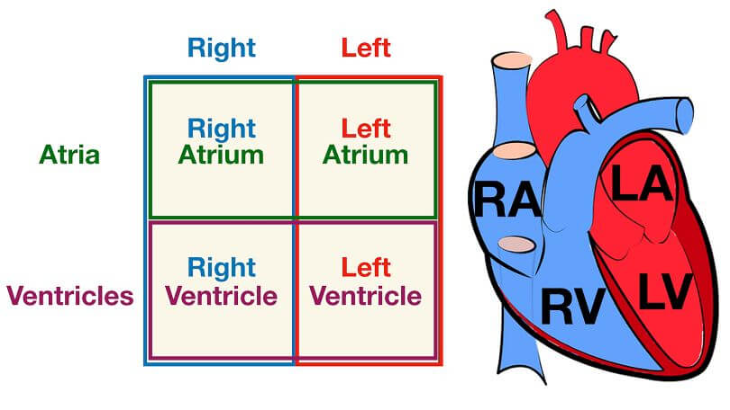
Four chambers make up the human heart. Because of this, humans are classified as four-chambered creatures, along with carnivores and other mammals. In the heart, the chambers are divided by the muscular septum. Each chamber of the heart has an interatrial and intraventricular septum that serves as a barrier between them.
Atrium Vs. Ventricle
The atrium and ventricle are two distinct parts of the heart of more sophisticated animals. Due to their position, the blood flow to and from the chamber is likewise different. The sizes and sorts of values that they include also vary.
Two of the heart’s four atria are referred to as the atriums. The interatrial septum separates the two atria in the human heart. The atrium receives deoxygenated blood from the body and sends it to the lungs, where it is reoxygenated and returned to the rest of the body. In comparison to the ventricles, it is smaller.
The lowest two chambers of the heart are referred to as the ventricles. The interventricular septum separates the two ventricles of the human heart. The dorsal aorta is the major artery that pumps blood throughout the body, and the job of the ventricles is to assist in this process. It dwarfs the atria in terms of size.
What is the Atrium?
The top part of the heart is referred to as the atrium, which is a kind of auricle. The interauricular or interatrial septum separates the two chambers of the human heart, resulting in two auricles. Two veins, the lower vena cava and the superior vena cava, deliver deoxygenated blood to the right auricle or atrium. Basically, they are veins. The pulmonary vein in the lungs delivers oxygenated blood to the left auricle.
It accepts blood that has been deoxygenated and uses the pulmonary circulation to re-oxygenate it. There are no long distances to travel between the heart and lungs, hence the auricles’ walls are comparatively thin. As a result, the auricles have a lower blood pressure than the ventricles.
In addition, it transports blood to the heart’s ventricles, where it is dispersed to all parts of the body. The tricuspid valve connects the right and left ventricles, whereas the mitral valve connects the left and right ventricles. The sinoatrial node and the pacemaker cells are located in the auricular area.
Sinoatrial node impulses control the heartbeat rate and rhythm. Because of this, the heart’s natural pacemaker is also known.
What is a ventricle?
Lower half of heart referred to as ventricles is known as a ventricle. The interventricular septum divides the heart’s lower chambers. The aorta transports blood from the right ventricle to the rest of the body through the left ventricle, which gets it from the auricles.
Various arteries and networks convey oxygenated cardiac blood to every organ of the body. Because of the great distance between the heart and the lower body or the kings, the blood pressure in this chamber is high and its walls are strong and muscular.
This is because the ventricle exerts a lot of pressure while pumping blood, which is why our pulse tends to lean slightly left. By way of the semilunar valve, the right ventricle enters the pulmonary artery, and by way of the aortic valve, the left ventricle enters the aorta. Both ventricles are connected through the heart.
It’s also larger as a result of the increased functionality and power it requires. Its Purkinje fibers carry electrical impulses across the ventricles of the brain.
Difference Between Atrium and Ventricle
- In comparison to the ventricles, the atrium is smaller and thinner.
- The heart and lungs are supplied by the atrium, while the whole body is supplied by the ventricles.
- Unlike the ventricles, the atrium is always filled with deoxygenated blood.
- Unlike the ventricles, the atrium’s walls are not as thick as those of the ventricles.
- The aorta drains the ventricles, while the aorta drains the atrium.
- The vena cava supplies the atrium, whereas the pulmonary vein supplies the ventricles.
- The atrium is responsible for maintaining the heart’s beat, whereas the ventricles are responsible for assisting it.
Conclusion
Uniform blood circulation requires the appropriate operation of each of the heart’s chambers. A shortage of blood flow to the heart may result from a blockage in any of the draining or supplying pathways.
In order to create the heart, the auricles and the ventricles must be put together. As previously mentioned, they are separated by septums yet linked by a tiny channel protected by valves. The emergence of the heart’s numerous chambers is a result of the heart’s ability to divide labor. Humans have a twofold circulatory system, and this method simplifies circulation.